|
Palomar College |
Physiological Psychology |
Dept of Behavioral Sciences |
|
|
Roger N. Morrissette, PhD |
|
Brain Dissection Lab
Objectives:
By the end of this laboratory you should be able to do the following:
1. Identify all regions of the dorsal surface of a sheep brain.
2. Identify all regions of the ventral surface of a sheep brain.
3. Identify all regions of the sheep brain following a midsagittal cut.
This laboratory involves a video dissection of a sheep brain and the comparison of that sheep brain to other animal brains. Please begin the lab by reviewing the lecture on Brain Evolution. Please print out these slides ahead of time and be prepared to take notes. The content of which will be on your next quiz or examination. Since this online laboratory involves dissection of animal tissue it is important to remember to be thankful, respectful, and subsequently take your studies seriously. The brain is the most complex and fascinating organ in an animal’s body. The anatomical structures that you see in the sheep brain are an extremely close approximation to those in your own brain. The regions are the same, the cranial nerves are the same, but fortunately for most of you, your brains are a bit larger and somewhat more complex than that of a sheep. You will find throughout this preparation that I have stated the functions of the individual brain region or nerve in italics and in parentheses just following that item. When you have completed the video of the sheep brain dissection, read the complete laboratory and answer the questions on the Review Sheet at the end of the laboratory.
Good Luck.
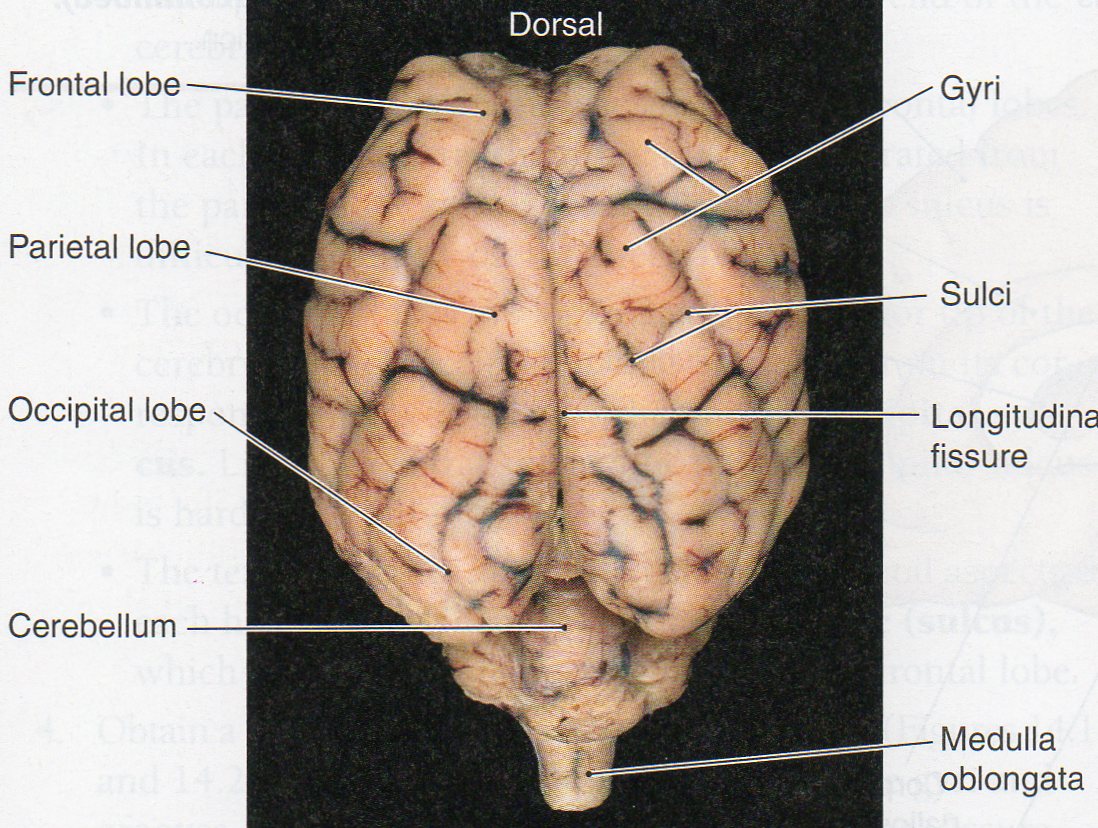
1. Identify all regions on the dorsal surface of a sheep brain.
We will begin our investigation with the dorsal surface of the intact sheep brain. Since the sheep is a 4-legged animal, the top of the skull is considered the back or dorsal side. The underside of the brain is called the ventral side. Remember from laboratory 1, we use the terms “rostral” and “caudal” to refer to the forehead side and spinal cord side of the brain respectively. We also use the terms “medial” and “lateral” to refer to "towards the midline" and "away from the midline" respectively. Use the figure below to identify the largest portion of the brain the Cerebrum (the entire cortical region area) and the Frontal (motor strip; strategy, reasoning, long-term planning), Parietal (somatosensory area), and Occipital (vision) lobes respectively. This region has many folds or gyri (gyrus – singular) and grooves or sulci (sulcus – singular). Identify the central groove the medial longitudinal fissure. This separates the right and left hemisphere of the cerebral cortex (integration of information, thought center). Moving caudally identify the Cerebellum (fine motor coordination) with its medial section, the Cerebellar Vermis and its two lateral sections, the Cerebellar Hemispheres. Also locate the Medulla (vital functions), and Spinal Cord (pathway of sensory and motor neurons).
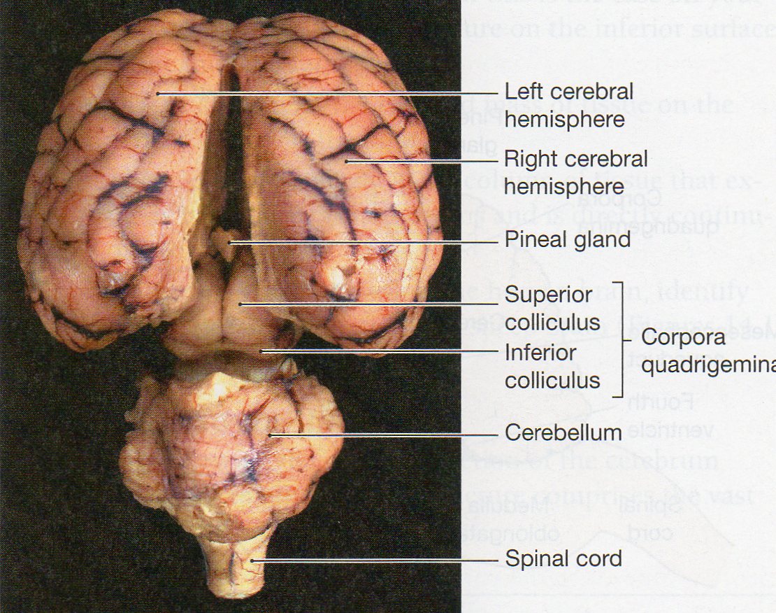
Now gently flex the brain between the Cerebellum and Cerebrum and peak inside to see the Midbrain or Mesencephalon (sleep/arousal regulation area) region which is made up of the Corpora Quadrigemina or even more specifically, the Superior Colliculus (visual information pathway) and the Inferior Colliculus (auditory information pathway). If you delicately remove the connective tissue that holds the Right and Left Cerebral Hemispheres together, you should be able to see the Pineal Gland (sleep and circadian rhythms).
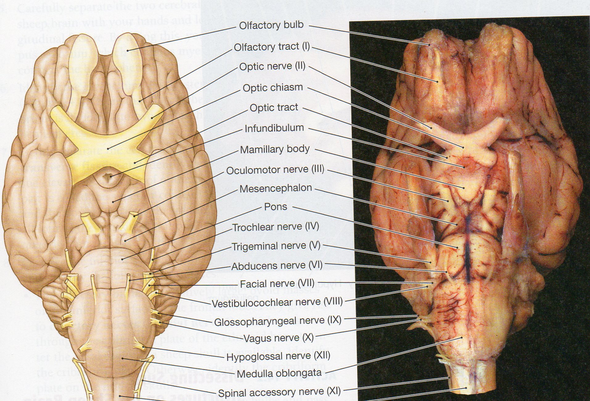
2. Identify all regions on the ventral surface of a sheep brain.
Now gently turn your sheep brain over to expose its’ ventral surface. The full Brainstem, all or most of the Cranial Nerves, and Temporal lobe (or Pyriform lobe) should be clearly visible. Use the figures below to identify the three regions of the brainstem: the Medulla, Pons (REM sleep area, nerve pathway) and Midbrain. Identify also the large lateral projection of the Temporal or Pyriform lobes (auditory center). Identify the medially placed Hypothalamus (center for lusts) and Pituitary Gland (if still attached). Your last and most difficult task is to identify as many of the 12 Cranial Nerves as you can. Nerves are bundles of axons that are either coming or going to a group of specific cells or nuclei. Start rostrally and identify the two large olfactory bulbs and nerves - cranial nerve I (detect smells), the optic nerve - II (visual information) and optic chiasm and optic tract, the oculomotor nerve – III (eye movement, pupil constriction), the trochlear nerve – IV (controls extra-ocular muscles), trigeminal – V (face senses and movement, mastication), abducens – VI (eye movement), facial – VII (facial senses, salivation), vestibulocochlear – VIII (balance and hearing), glossopharyngeal – IX (taste), vagus – X (visceral system regulator), accessory – XI (neck and shoulder muscles), and hypoglossal nerve – XII (tongue movement). These nerves can be very hard to find. You might want to try to clean up your brains by gently removing the meningial coverings and any blood vessels that are in the way. Blood vessels should be dark while all of the myelin coated nerves will be white. Take your time, use the diagrams below, move around gently and you should be able to find most if not all of the cranial nerves.
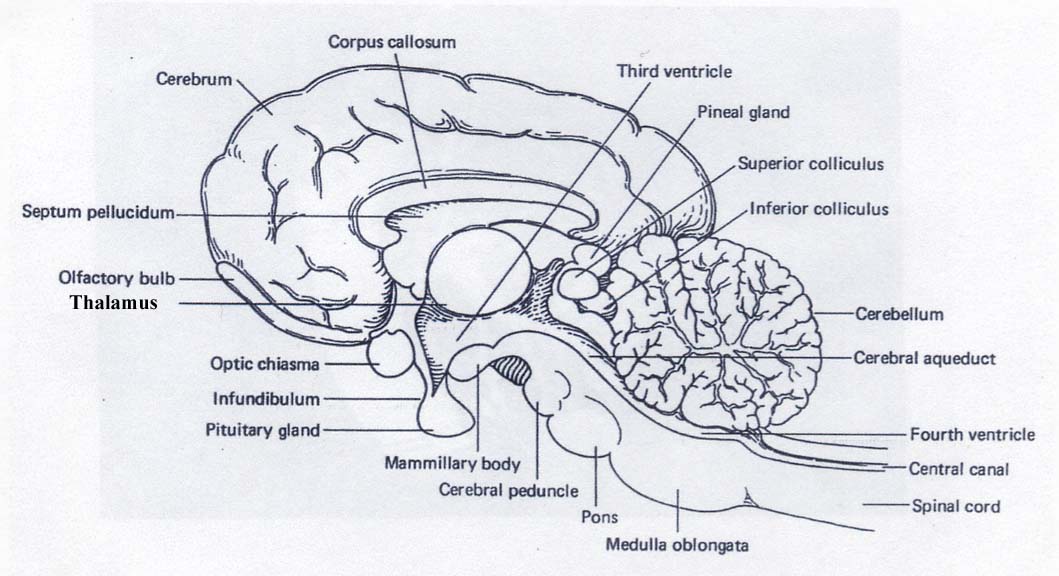
3. Identify all regions of the sheep brain from a midsagittal section.
For this final view, use the scalpel and cut your brain in half making a midsagittal slice. Observe one half of the brain and compare it to the labeled figure below. Identify the Cerebrum, Corpus Callosum (pathway connecting right and left hemispheres), Septum Pellucidum (separates the lateral ventricles), Optic Chiasm, Thalamus (great relay station), Pineal Gland (sleep and circadian rhythms), Superior Colliculus (visual information relay pathway), Inferior Colliculus (auditory information relay pathway), Hypothalamus, Midbrain, Pons, Medulla, Cerebellum, and the Spinal Cord.

Human Brain
Review Sheet
Exercise 1
Identify each region in the midsagittal section below

1. ________________________________ 8. ________________________________
2. ________________________________ 9. ________________________________
3. ________________________________ 10. ________________________________
4. ________________________________ 11. ________________________________
5. ________________________________ 12. ________________________________
6. ________________________________ 13. ________________________________
7. ________________________________ 14. ________________________________
Exercise 2
Place the number of the appropriate region next to its’ appropriate function
Region or Nerve Function
1. cerebral cortex ______ center for lusts
2. hypothalamus ______ circadian rhythms
3. medulla ______ taste
4. pons ______ detect smells
5. thalamus ______ balance and hearing
6. corpus callosum ______ tongue movement
7. CN I ______ auditory information relay pathway
8. CN II ______ integration of information, thought center
9. CN III ______ REM sleep area, nerve pathway
10. CN IV ______ facial senses, salivation
11. CN V ______ neck and shoulder muscles
12. CN VI ______ visual information
13. CN VII ______ fine motor coordination
14. CN VIII ______ great relay station
15. CN IX ______ pathway connecting right and left hemispheres
16. CN X ______ auditory center
17. CN XI ______ sleep/arousal regulation area
18. CN XII ______ eye movement
19. cerebellum ______ visual information relay pathway
20. spinal cord ______ separates the lateral ventricles
21. midbrain ______ pathway of sensory and motor neurons
22. temporal lobe ______ vital functions
23. septum pellucidum ______ face senses and movement, mastication
24. superior colliculus ______ eye movement, pupil constriction
25. inferior colliculus ______ controls extra-ocular muscles
26. pineal gland ______ visceral system regulator
Please scan or take a photo of your Review Sheet in hand it in by the deadline to get credit for this laboratory.