|
Palomar College |
Physiological
Psychology |
DAY COURSE |
|
|
Roger N. Morrissette, PhD
|
|
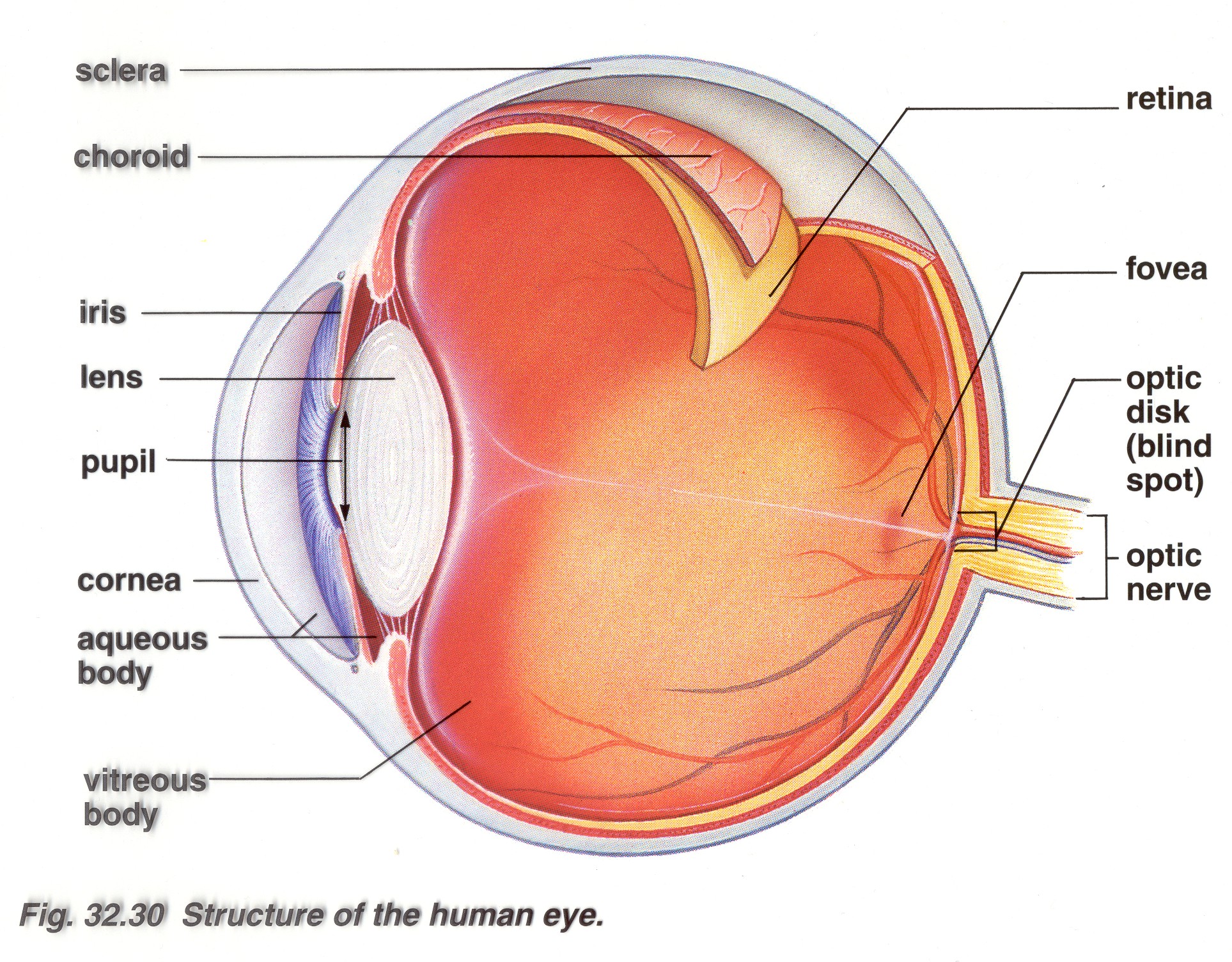
Visual System
Objectives:
By the end of this laboratory you should be able to understand
the following concepts associated with the:
Human Visual Sensory System:
By the end of this laboratory you should be able to do the following:
-
Successfully dissect, sketch, and identify the
superficial aspect of the eyeball.
-
Successfully dissect, sketch, and identify the
deep aspect of the eyeball.
-
Successfully dissect, sketch, and identify the
lens.
This
laboratory investigates the many facets of the sensory system. We will begin the lab with a lecture on
The Visual System.
Please print out these slides ahead of time and be prepared to take notes. The
content of which will be on your next quiz or examination.
The body’s homeostatic process
begins with its sensory receptors. These receptors monitor many aspects of
the internal and external environment and relay that information to the nervous
system. The body uses that information to adequately respond to the
environment. Sensory receptors come in many types: Mechanoreceptors interpret physical deformation of
different body parts; Chemoreceptors monitor
different chemical concentrations; Photoreceptors
record light energy; and Thermoreceptors send
information about temperature to the brain. In today’s laboratory we will
be conducting exercises with a specific sensory system -
The Visual System. Please choose a single partner to
work with and read the exercise directions carefully. Please also take excellent
notes of your findings so that I can check them at the end of the class.
Good luck.
Human
Visual Sensory System
Blind Spot Detection:
To measure your blind spot you will need to use the small strip of
paper that has both a black dot and a black cross on it.
1. Hold the paper in your right hand with the black dot on the right
side of the cross.
2. Fully extend your right arm and position the paper so that it is
at your eye level.
3. Close your left eye and focus your right eye only on the cross.
4. Now very slowly bring the
paper closer to your eyeball.
5. At a certain distance, you will see the spot disappear from your
field of vision - that is your blind spot.
6. At that point, have your partner measure the distance between your
eye and the paper.
Blind Spot ________________
from the eye.
Dominant Eye Determination:
Most people do not use their two eyes equally. Typically one eye is more
dominant than the other.
To determine which is your dominant eye, start by:
1. Rolling up an 8 1/2" x 11" sheet of paper into a long tube about 4 cm
in diameter.
2. Hold the tube at arms length between both of your eyes and notice the
image you see through the tube.
3. Now close one eye and then the other. Your dominant eye will see the
same image as the one both of your eyes sees.
Which eye is your dominant eye?
_____________________
What is your dominant hand?
___________________
Visual Accommodation Exercises
Proper
focusing on objects of varying distances requires the eye to
accommodate by adjusting both the lens shape and the size of the
opening. In the following exercises you will observe some
conditions under which these automatic accommodations take place.
Near Point of Vision:
The distance from the eye to the nearest object that can be focused
clearly is called the near point of vision. To identify your near point of
vision for each of your eyes you must first:
1. Pick up a straight pin with your right hand and hold it vertically at
arm's length and at the level of your eyes.
2. Place your left hand over your left eye.
3. Gradually bring the pin closer to your eye, focusing continually on
the pin until it begins to blur.
4. Have your partner measure the distance between your eye and the point
where the pin began to blur - this is your near point vision.
What is your near point vision
for your right eye? _____________________
5. Repeat the procedure with your left eye.
What is your near point vision
for your left eye? _____________________
For this exercise you will be observing your partners pupils. If they
have a light colored iris this should be pretty easy, but if they have a
dark colored iris you may have to get pretty close to see the pupil
changes.
1. Grab a penlight and turn it on.
2. Look closely at the shape of your partners right eye pupil.
3. Now quickly flash the penlight over and away from your partners right
eye.
What do you observe about their
pupil? _______________________________
4. Repeat the procedure for their left eye.
Can you observe any difference in
the response? _________________________________
5. Take another close look at your partner's right eye pupil.
6. While still observing your partner's right eye pupil, have them place
their hand over their left eye for 1 minute and then remove it.
What do you observe about their
pupil? _______________________________
7. Repeat the procedure for their left eye.
Can you observe any
difference in the response? _________________________________
Today’s laboratory
also involves the dissection of a sheep
eye. Since this laboratory involves dissection of animal tissue it is important
to remember to be thankful, respectful, and subsequently take your studies, and
in this case, your dissections seriously. Once again you will be using extremely
sharp scalpels and probes so be careful with them. Please proceed with caution
and care during your dissections. Work in groups of no more than two. Use the
dull and sharp probes to try to identify all of the brain regions discussed.
Follow the instructions carefully before you cut into your eyeballs. A single
clean slice will make viewing clearer. I will be asking you to identify the
different regions and parts of your sheep eyes so study while you dissect. When
you have completed your dissection you will need to do the exercises on the last
pages of the handout (which will be given to you in class) in order to complete
the laboratory. Good Luck in your explorations.
Successfully dissect, sketch, and identify the
superficial aspect of the eyeball.
|
We receive the majority of our
information through our visual senses. There is much integration of the
images by our retina prior to getting to our visual cortex. The rods are
critical for dim light discrimination and periphery viewing. The cones are
located near the fovea and are important for color discrimination and fine
details. Both the left and right eye work together to give you a clear
view of the world. These exercises should help you gain a better respect
for your visual system. |
 |
Step
1: Trim away excess adipose tissue (yellow material), the extrinsic eye
muscles (tan material), and the connective (conjunctiva) tissue (white/clear
material). If you pull with the tweezers you will see the clear connective
tissue holding the muscle and fat in place. In this taught position you can
simply cut all of the muscle and fat off of the eyeball.
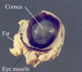
Step 2: Draw and color a side
view of your eye specimen. Label all of the listed terms.
|
optic nerve
sclera
cornea
pupil
conjunctiva
|
Step 3: Give the function of the
five terms you identified.
optic nerve: _________________________________
sclera: _____________________________________
cornea: ____________________________________
pupil: ______________________________________
conjunctiva: _________________________________
Successfully dissect, sketch, and identify the
deep aspect of the eyeball.
|
Step 1:
Puncture the sclera with the tip of the scissors or pointer and cut a
circle parallel with the cornea about 1 cm beyond the edge of the cornea
and sclera. See the diagram to the right for details. |
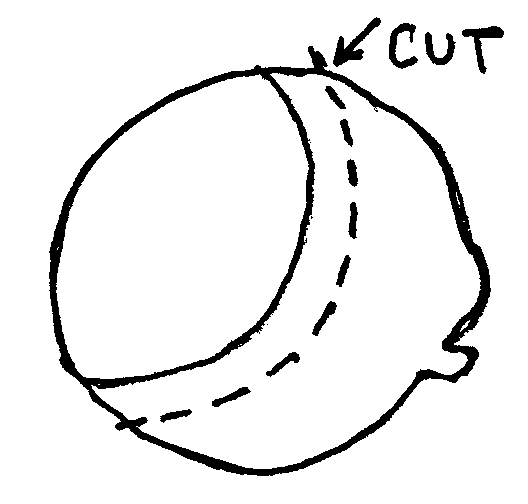 |
Step 2: Gently tease apart the
anterior and posterior portions of the eyeball. The fluid that escapes is the
aqueous humor. Be careful not to disturb the fine tissue layer in the posterior
section. That is the retina.
Step 3: Draw and color the
anterior view of your eye specimen. Label all of the listed terms.
|
aqueous humor
lens
iris
cilliary muscles
pupil
cornea
|
Step 4: Give the function of the
four undefined terms you identified.
aqueous humor: _______________________________________
iris: _________________________________________________
lens: _________________________________________________
cilliary muscles:
________________________________________
Step 5: Draw and color the
posterior view of your eye specimen. Label all of the listed terms.
|
vitreous
humor
retina
blind spot
tapetum
fovea
choroid coat
|
Step 6: Give the function of the
six terms you identified.
vitreous humor: _________________________________
retina: _________________________________________
blind spot: ______________________________________
tapetum: _______________________________________
fovea: _________________________________________
choroid coat:
____________________________________
Successfully dissect, sketch, and identify the
lens.
Dissect out the lens so it is separate from all other tissue.
Use the scalpel to cleanly bisect the lens. Draw a sketch below of what you
find.
rmorrissette@palomar.edu